


Arthrosis can affect any joint, while degenerative-dystrophic lesions can be isolated or occur in multiple joints at once.
This pathology is not life-threatening, but significantly reduces its quality. Severe pain, disability increases over time, leading to disability.
In the early stages, conservative therapy is prescribed to help stop the disease from developing.
It is difficult to completely cure osteoarthritis deformans (DOA), but bone junction functionality can be preserved. In the later stages, only surgical treatment will help.
Development mechanism
Many have heard of a disease like arthrosis, but not everyone understands what it is. To do this, you need to understand how the joint works.
The surface of the bones that make up the joints is covered with smooth, slippery, flexible cartilage that cushions and protects from injury. In arthrosis, the blood supply to this area is cut off and the hyaline cartilage starts to deteriorate. Furthermore, degenerative-dystrophic changes occur in the capsule, ligaments, periarticular muscles, and other segments of articulation.
Usually, the disease develops slowly, but the pathological process can be accelerated by external factors. Much depends on the characteristics of the patient's body, co-morbidities and lifestyle.
Osteoarthritis develops as follows:
- In a certain area of the cartilage lining of the joint, the bloodstream is damaged and then deficient in nutrients. As a result of traumatic factors, the area of destruction increases.
- The body replaces the defects in the cartilage layer of the joints with mineralized tissue that has no pure structure.
- Gradually, abnormal growths (osteophytes) appear on the hyaline coating.
- In the background of the pathological process, healthy areas of cartilage are exposed to excessive stress. As a result, the work of the joint is interrupted and its surfaces are gradually destroyed.
Carefully!Arthrosis causes the destruction of joint bone surfaces, inflammation of the joint membrane, and compaction of the joint capsule. The joint gap narrows, the joint deforms, breaks down, then the patient may become disabled. Therefore, it is very important to diagnose the pathology in time and start treating it.
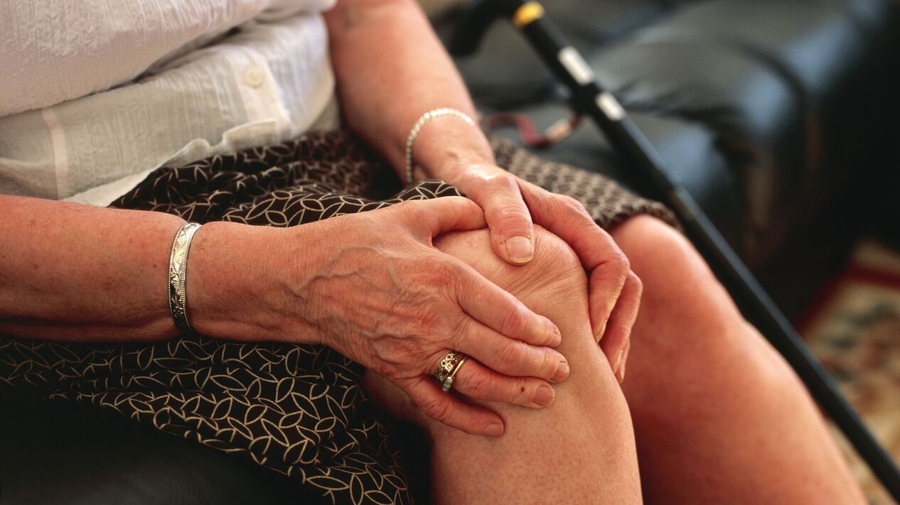
Arthrosis is usually seen in patients over 60 years of age. However, the disease is also diagnosed at a young age - between 20 and 45 years.
Reference. Arthritis and arthrosis are similar, so many patients are interested in the question of how the first disease differs from the second. In the case of DOA, only the joints are damaged, and arthritis is characterized not only by inflammation of the bone junction but also of the internal organs (liver, kidneys, heart). This is the main difference between these pathologies.
Classification
People who are far away from medicine when they hear names like gonarthrosis, coxoarthrosis, osteoarthritis do not understand the difference. The fact that there are many types of this pathology, which differ in their localization, characteristics, causes and origins. Therefore, physicians have developed several classifications of arthrosis to facilitate discrimination.
Types of arthrosis by localization:
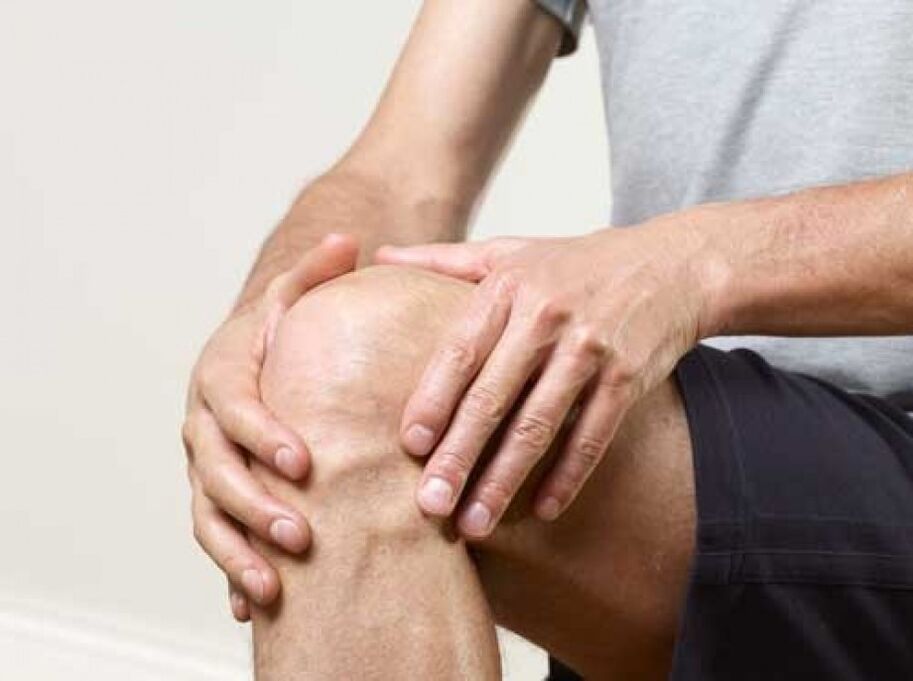
- Gonarthrosis is an abnormal process in the knee.
- Coxarthrosis is a change in the hip joint.
- Uncovertebral - deformity of the cervical spine.
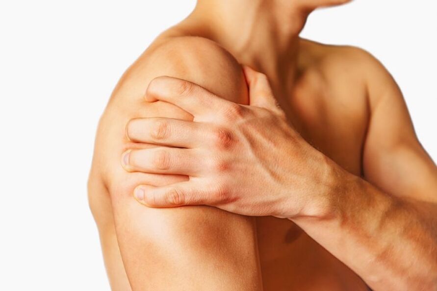
- Dystrophic changes in the shoulder joint.
- Interphalangeal - deformity of the interphalangeal joints of the bones.
- Spondyloarthrosis is a degenerative change in the joints of the spine.
- Ankle - Wear on the ankle joint.
- Polyosteoarthritis is a multiple disorder of the joints of the fingers.
In addition, there are jaw, temporal, cost-spine, clavicular-acromial arthrosis.
Depending on the characteristics of the course, the following pathologies are distinguished:
- Deforming arthrosis is a disease that has progressed to stage 3. It is a progressive disease that requires immediate surgery.
- Arthrosis-arthritis - destruction of cartilage lining, inflammation.
- Acute disease in which the characteristic symptoms become more pronounced.
- Chronic arthrosis is the slow destruction and thinning of the cartilage mucosa with a ruptured course.
Depending on the cause, the following are distinguished:
- It occurs due to dystrophic osteoarthritis - metabolic disorders.
- Fracture arthrosis - develops as a result of fracture.
- Post-traumatic - the disease appeared after a joint injury.
DOA varieties by origin:
- Primary (idiopathic) - occurs for no apparent reason, often due to age-related changes in bone joints.
- Secondary - degenerative-dystrophic disorders provoke a number of factors (metabolic disorders, hormonal imbalances, trauma).
Doctors distinguish between monoarthrosis and polyarthrosis. In the first case, 1 joint is affected, in the second, all joints are destroyed simultaneously. The last type of disease is called generalized arthrosis, in which 3 or more bone joints are deformed.
Pathological education
According to the symptoms and progression, 4 stages of DOA are distinguished:
- 1 degree.The shape and structure of the joint have not yet changed, so the disease is latent. The patient occasionally experiences mild discomfort in the affected area, especially after excessive physical exertion or sudden movements. The composition of the synovial fluid changes and the blood supply to the joint is disrupted. The muscle fibers surrounding the joint weaken.
- Grade 2.Bone joints begin to collapse, and bone growths form on their surface. Moderate painful feelings appear, inflammation occurs intermittently. During movement, a characteristic crackling is heard in the affected joint. Muscle functionality is impaired due to disruption of nerve tissue trophism.
- 3 degrees.Hyaline cartilage and articulation have marked degenerative disorders, causing the axis of the limb to bend. The ligaments and muscles are shortened, the joint becomes pathologically movable, but the movements are significantly limited. The patient often has incomplete dislocations.
- 4 degrees.The bone connection is completely destroyed, complete immobility is observed, and severe pain syndrome is observed even at rest.
Important. In the last stage of arthrosis, only the endoprosthesis helps (replacement of the affected joint with a prosthesis).
Causes of DOA
The question of why the disease occurs is quite relevant. Doctors distinguish between the internal (certain diseases, bad habits, unhealthy eating) and external (injuries, characteristics of professional activity) causes of osteoarthritis.
Often, secondary degenerative-dystrophic disorders develop in the background of the inflammatory process:
- Infectious diseases that provoke various viruses and bacteria.
- Rheumatism.
- Autoimmune disease.
- Purulent inflammation of the joint.
- Gout (deposition of uric acid salts on bone surfaces).
- Psoriasis of the joints.
DOA can occur due to cartilage abnormalities and malnutrition. The pathological changes are caused by the following factors:
- Genetic disorders.
- Pathologies occurring during intrauterine development.
- Age-related changes in the body.
- Osteoporosis (increased fragility of the bones due to calcium deficiency).
- Hormonal imbalance.
- Disorders of metabolic processes.
- Lack of vitamins and minerals.
- Pathologies accompanied by muscle weakness.
- Prolonged poisoning.
Exacerbation of diseases of the musculoskeletal system also provokes degenerative changes in cartilage tissue.
External factors in the development of arthrosis include:
- Regular hypothermia.
- Dislocations.
- High force on the articulation area.
- Fracture.
- Damage to the meniscus.
- Excessive physical activity related to professional sports or professional activities.
- Obesity.
- Surgery of joints or periarticular structures.
Regardless of the causes of DOA, it is important to first identify the cause of the abnormal lesions and then address the consequences.
Reference.Idiopathic arthrosis occurs on its own for no apparent reason.
Symptoms
Arthrosis manifests itself in the following symptoms:
- pain syndrome;
- restrictions on mobility;
- cracking while moving;
- edema, a change in the axis of the relationship.
These are typical symptoms that occur in all patients.

The initial signs of pathology are discomfort in the affected area that occurs after physical exertion. After the onset of discomfort, a doctor should be consulted as it can be cured in the early stages of the disease.
Later, the patient complains of mild to moderate pain that appears after the injured joint is loaded and disappears quickly.
Decreased bone junction mobility indicates degenerative changes in its structure. At first, the patient feels stiff, especially in the morning. Later, it becomes increasingly difficult for the patient to perform active movements. With further development, mobility restrictions are even more helpful. If left untreated, joint contracture occurs and over time, motor activity is blocked.
Many patients complain of cracks in the joints while moving, accompanied by painful feelings and decreased mobility. As the disease progresses, this manifestation becomes more pronounced.
In later stages, the axis of the limb bends and the joint membrane deforms. This indicates that the bone connection has virtually collapsed and healthy tissues have been replaced by osteophytes. At this stage, adjacent joints are subjected to heavy stress, thus increasing the likelihood of damage to their cartilage lining.
Establishing the diagnosis
If you notice signs of osteoarthritis, see a doctor immediately. The diagnosis of DOA is made after a medical history, laboratory and instrumental examinations.
First, a visual examination is performed, during which the doctor may notice swelling in the affected area. In addition, a palpation is performed that allows the determination of pain, lumps, temperature changes, and skin moisture.
Comprehensive diagnostics include laboratory research. Blood tests may show an inflammatory process indicated by an increase in ESR (erythrocyte sedimentation rate), an increase in uric acid concentration. A urine test is performed to determine protein levels.
Arthrosis is diagnosed by the following instrumental tests:
- X-rays help to see the change in the shape of the joint.
- Contrast-enhanced arthrography is a more accurate diagnostic method than X-rays.
- CT is used to assess the structure of the joint.
- Radionuclide diagnostics are performed using radiopharmaceuticals. This study makes it possible to assess the anatomical and functional state of the relationship.
Magnetic resonance imaging is a modern, highly informative diagnostic method. During the examination, deformation of the damaged joint, rupture of the meniscus or ligament can be seen.
To examine the synovial fluid, doctors prescribe a puncture of the affected limb.
After diagnostic procedures, the doctor will establish a treatment regimen.
Healing methods
Medical treatment is required for arthrosis of any stage. Complex therapy performed at an early stage helps to stop pathological changes and restore joint function. If the patient visits a doctor in the late stages of the DOA, the prognosis is poor.
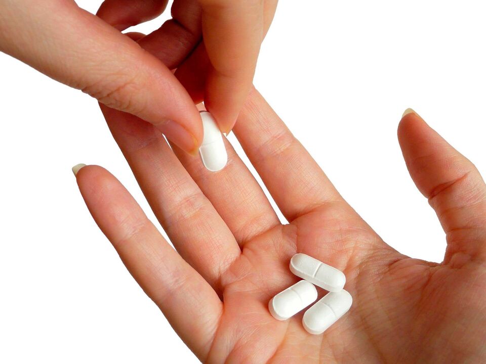
medication is used for grade 2 arthrosis. Chondroprotectors are used to restore the structure of the cartilage. The patient is prescribed medications in the form of tablets and capsules. It should be taken twice a year in 3-4 month courses. The drug contains the structural elements of the cartilage lining.
Non-steroidal anti-inflammatory drugs (tablets, injections) help relieve pain.
DOA treatment is performed by physiotherapy methods:
- Magnetic therapy.
- Ultra-high frequency therapy.
- Electrophoresis.
- Shockwave therapy.
- Paraffin therapy.
- Sludge treatment.
Physiotherapy and physiotherapy exercises are performed after the pain has disappeared. The physician compiles a series of exercises that the patient should perform systematically. Exercise therapy increases muscle tone, strengthens ligaments, normalizes blood circulation and helps restore articulation.
It is recommended to provide rest during and after the treatment, to reduce the load on the patient's joint with the help of bandages, crutches and sticks.
Sometimes the patient prescribes a massage. After a procedure, the blood supply to the affected area improves and the pain decreases.
During therapy, the patient must eat properly. Sugar, flour, fatty, spicy foods, potatoes, tomatoes, eggplant should be given up. And it is recommended to get rid of bad habits (alcohol, smoking) forever.
Intra-articular injections are used for arthrosis:
- Glucocorticosteroids help to normalize the blood supply to the affected area, stop the inflammatory process, increase the elasticity of bone tissue.
- Joint fluid analogs with chondroprotective properties. These drugs reduce pain, improve joint mobility, and accelerate the production of collagen and elastane.
In the last stage of the DOA, surgical treatment methods are used:
- Endoprosthesis.
- Arthrodesis.
- Arthroscopy.
In advanced cases, doctors replace the destroyed compound with a metal prosthesis. This method is most commonly used to treat large joints. After surgery, the patient's quality of life improves.
If arthrodesis cannot be performed, the patient is prescribed arthrodesis. During surgery, the bone surfaces are fixed with a special plate. Arthrodesis helps relieve severe pain but does not restore joint motor activity.
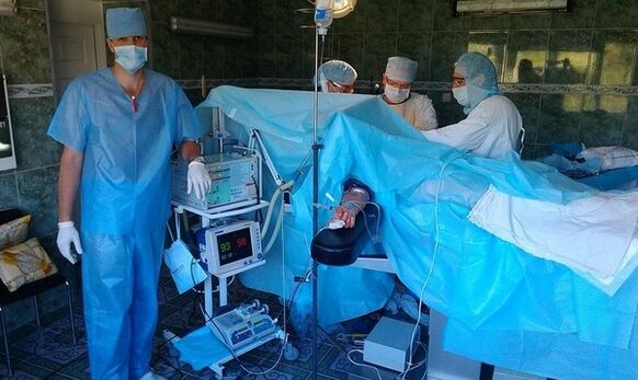
During arthroscopy, a miniature camera and manipulators are inserted into the joint cavity to help remove bone growths and restore cartilage structure. The camcorder allows you to monitor all the manipulations on the screen. The operation is usually performed in gonoarthrosis, but its effect lasts for a short time.
DOA is dangerous, so it is important to identify and manage it in a timely manner.
Opinions
Patients with arthrosis find it easiest to cure in the early stages of the disease. In advanced cases, only surgery will help. But in both cases, the treatment must be comprehensive.
- The first opinion: "I was diagnosed with grade 2 knee arthrosis 1 year ago. I took special medications, participated in physiotherapy, and dieted. At first, the pain was gone, mobility was restored, but after 3-4 months, the symptoms returned again. Sometimes the pain was accompanied by a rise in temperature. The doctor advised me to take capsules with hondoprotectors. With them, my condition improved, I didn’t feel any pain for six months. "
- Second Review: "A few years ago, I was diagnosed with grade 3 coxoarthrosis. It kept hurting, even at night, I couldn’t move my leg normally. The doctor advised me to have surgery, but at first I refused and decided to try intraarticular injections. However, after the procedures, my condition did not change much. As a result, I opted for a radical method. He recovered for 1 year to 3 months after endoprosthesis. During this period, he took medication, performed special exercises, went to massages, physiotherapy, and followed a diet. I live a full life now. I advise everyone not to hesitate with the treatment. "
- Third Review: "After MRI, I was diagnosed with rupture of the internal meniscus of the knee and grade 1 gonoarthrosis. Doctors prescribed chondroprotectors. I used the ointment twice a day. I used an orthosis to protect my knee, I just took it off at home while resting. After the injections, the electrophoresis and the paraffin therapy started, I also bought a magnetic therapy device, it was 10 times. After another diagnosis, doctors said the joint had recovered in 70%. I will continue the treatment and hope to fully recover my legs. "
As you can see, there are different types of arthrosis. Medical help should be sought at the first suspicious signs to avoid surgery and restore joint function. Only a doctor can determine the type of disease, the degree of its complexity, and make an appropriate treatment regimen. DOA is easier to manage early on.